
Page 4 - wjols
P. 4
Ten-point Strategy for Safe Laparoscopic Cholecystectomy
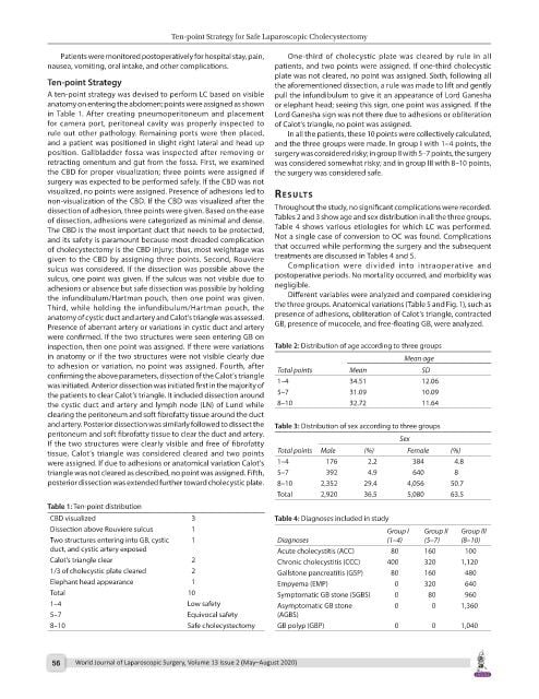
Patients were monitored postoperatively for hospital stay, pain, One-third of cholecystic plate was cleared by rule in all
nausea, vomiting, oral intake, and other complications. patients, and two points were assigned. If one-third cholecystic
plate was not cleared, no point was assigned. Sixth, following all
Ten-point Strategy the aforementioned dissection, a rule was made to lift and gently
A ten-point strategy was devised to perform LC based on visible pull the infundibulum to give it an appearance of Lord Ganesha
anatomy on entering the abdomen; points were assigned as shown or elephant head; seeing this sign, one point was assigned. If the
in Table 1. After creating pneumoperitoneum and placement Lord Ganesha sign was not there due to adhesions or obliteration
for camera port, peritoneal cavity was properly inspected to of Calot’s triangle, no point was assigned.
rule out other pathology. Remaining ports were then placed, In all the patients, these 10 points were collectively calculated,
and a patient was positioned in slight right lateral and head up and the three groups were made. In group I with 1–4 points, the
position. Gallbladder fossa was inspected after removing or surgery was considered risky; in group II with 5–7 points, the surgery
retracting omentum and gut from the fossa. First, we examined was considered somewhat risky; and in group III with 8–10 points,
the CBD for proper visualization; three points were assigned if the surgery was considered safe.
surgery was expected to be performed safely. If the CBD was not
visualized, no points were assigned. Presence of adhesions led to results
non-visualization of the CBD. If the CBD was visualized after the
dissection of adhesion, three points were given. Based on the ease Throughout the study, no significant complications were recorded.
of dissection, adhesions were categorized as minimal and dense. Tables 2 and 3 show age and sex distribution in all the three groups.
The CBD is the most important duct that needs to be protected, Table 4 shows various etiologies for which LC was performed.
and its safety is paramount because most dreaded complication Not a single case of conversion to OC was found. Complications
of cholecystectomy is the CBD injury; thus, most weightage was that occurred while performing the surgery and the subsequent
given to the CBD by assigning three points. Second, Rouviere treatments are discussed in Tables 4 and 5.
sulcus was considered. If the dissection was possible above the Complication were divided into intraoperative and
sulcus, one point was given. If the sulcus was not visible due to postoperative periods. No mortality occurred, and morbidity was
adhesions or absence but safe dissection was possible by holding negligible.
the infundibulum/Hartman pouch, then one point was given. Different variables were analyzed and compared considering
Third, while holding the infundibulum/Hartman pouch, the the three groups. Anatomical variations (Table 5 and Fig. 1), such as
anatomy of cystic duct and artery and Calot’s triangle was assessed. presence of adhesions, obliteration of Calot’s triangle, contracted
Presence of aberrant artery or variations in cystic duct and artery GB, presence of mucocele, and free-floating GB, were analyzed.
were confirmed. If the two structures were seen entering GB on
inspection, then one point was assigned. If there were variations Table 2: Distribution of age according to three groups
in anatomy or if the two structures were not visible clearly due Mean age
to adhesion or variation, no point was assigned. Fourth, after Total points Mean SD
confirming the above parameters, dissection of the Calot’s triangle
was initiated. Anterior dissection was initiated first in the majority of 1–4 34.51 12.06
the patients to clear Calot’s triangle. It included dissection around 5–7 31.09 10.09
the cystic duct and artery and lymph node (LN) of Lund while 8–10 32.72 11.64
clearing the peritoneum and soft fibrofatty tissue around the duct
and artery. Posterior dissection was similarly followed to dissect the Table 3: Distribution of sex according to three groups
peritoneum and soft fibrofatty tissue to clear the duct and artery.
If the two structures were clearly visible and free of fibrofatty Sex
tissue, Calot’s triangle was considered cleared and two points Total points Male (%) Female (%)
were assigned. If due to adhesions or anatomical variation Calot’s 1–4 176 2.2 384 4.8
triangle was not cleared as described, no point was assigned. Fifth, 5–7 392 4.9 640 8
posterior dissection was extended further toward cholecystic plate. 8–10 2,352 29.4 4,056 50.7
Total 2,920 36.5 5,080 63.5
Table 1: Ten-point distribution
CBD visualized 3 Table 4: Diagnoses included in study
Dissection above Rouviere sulcus 1 Group I Group II Group III
Two structures entering into GB, cystic 1 Diagnoses (1–4) (5–7) (8–10)
duct, and cystic artery exposed Acute cholecystitis (ACC) 80 160 100
Calot’s triangle clear 2 Chronic cholecystitis (CCC) 400 320 1,120
1/3 of cholecystic plate cleared 2 Gallstone pancreatitis (GSP) 80 160 480
Elephant head appearance 1 Empyema (EMP) 0 320 640
Total 10 Symptomatic GB stone (SGBS) 0 80 960
1–4 Low safety Asymptomatic GB stone 0 0 1,360
5–7 Equivocal safety (AGBS)
8–10 Safe cholecystectomy GB polyp (GBP) 0 0 1,040
56 World Journal of Laparoscopic Surgery, Volume 13 Issue 2 (May–August 2020)