
Page 39 - wjols
P. 39
Total Laparoscopic Pancreaticoduodenectomy
Table 1: Frequency of pancreaticoenteric and pancreaticogastric
anastomosis
Anastomosis Frequency
Pancreaticojejunal (dunking) 8
Pancreaticojejucal (duct-to-mucosa) 14
Pancreaticogastric (dunking) 11
anastomosis. The site for pancreaticogastrostomy was marked
higher on the body of the stomach and anterior gastrotomy was
performed opposite to it. The pancreatic stump was brought inside
a smaller posterior gastrotomy so as to have a snug placement
of pancreas inside stomach which was sutured with continuous
sutures with 2.0 silk leaving at least 1 cm of pancreatic stump inside
the stomach. The anterior gastrotomy was closed with 2.0 silk in two
layers (Table 1). Gastrojejunal anastomosis was performed with 3.0
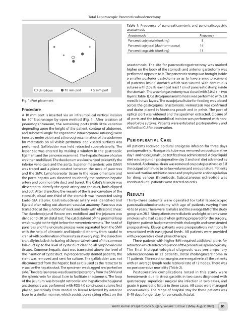
Fig. 1: Port placement mersilk in two layers. The nasojejunal tube for feeding was placed
across the gastrojejunal anastomosis. Hemostasis was confirmed
Procedure and drains placed in Morrisons pouch and in pelvis. The port of
A 10 mm port is inserted via an infraumbilical vertical incision optical port was widened and the specimen extracted. Closure of
for 30° laparoscope by open method (Fig. 1). After creation of all ports and the infraumbilical incision was performed with non-
pneumoperitoneum, the remaining ports (with little variation absorbable sutures. Patients were extubated postoperatively and
depending upon the height of the patient, contour of abdomen, shifted to ICU for observation.
and subcostal angle for ergonomic intracorporeal suturing) were
inserted under vision and a thorough examination of the abdomen perIoperAtIve cAre
for metastasis on all visible peritoneal and visceral surfaces was
performed. Gallbladder was held retracted superolaterally. The All patients received epidural analgesia infusion for three days
lesser sac was entered by making a window in the gastrocolic postoperatively. Nasogastric tube was removed on postoperative
ligament and the pancreas examined. The hepatic flexure of colon day 1 and nasojejunal tube test feed was administered. A clear liquid
was then mobilized. The duodenum was kocherized to identify the diet was begun on postoperative day 3 and oral diet advanced as
inferior vena cava and the aorta. Superior mesenteric vein (SMV) tolerated. Abdominal drain was removed on postoperative day 5 if
was traced and a plane created between the neck of pancreas the output continued to be low volume and serous nature. Patients
and the SMV. Lymphoareolar tissue in the lesser omentum and received routine antibiotic cover and prophylactic anticoagulation
the porta hepatis was dissected to identify the common hepatic for deep venous thrombosis. Subcutaneous octreotide was
artery and common bile duct and bared. The Calot’s triangle was continued until patients were started on orals.
dissected to identify the cystic artery and the duct, both clipped
and cut. After dissecting the vessels of the lesser curvature of the
stomach, distal one-third of the stomach was transected using results
Endo-GIA stapler. Gastroduodenal artery was identified and Thirty-three patients were operated for total laparoscopic
ligated after ruling out aberrant vascular anatomy. Pancreas was pancreaticoduodenectomy with age of patients varying from
transected at the junction of neck and body with ultrasonic shear. 45 to 67 years. There were 13 males and the average BMI of the study
The duodenojejunal flexure was mobilized and the jejunum was group was 28.3. Nine patients were diabetic and eight patients were
divided 10–20 cm distal to it. The cut distal end of the proximal loop smokers who had ceased when getting prepared for the surgery.
was brought to the right below the mesenteric vessels. The head of Eighteen patients had presented with cholangitis and were stented
pancreas and the uncinate process were separated from the SMV preoperatively. Eleven patients were preoperatively nutritionally
with the help of ultrasonic and bipolar diathermy from caudal to resuscitated with nasojejunal feeds. All patients were provided
cranial with confirmation of hemostasis at every step. The dissection with preoperative chest physiotherapy.
cranially included the baring of the portal vein and of the common Three patients with higher BMI required additional ports for
bile duct up to the level of cystic duct clearing all lymphovascular retraction which aided completion of the procedure laparoscopically.
tissues. Common hepatic duct was transected above the level of The final histopathological diagnosis was periampullary
the insertion of cystic duct. In preoperatively stented patients, the adenocarcinoma in 22 patients, distal cholangiocarcinoma in
stent was removed and sent for culture. The gallbladder was not 11 patients. The resection margins were negative in all the patients
disconnected from the hepatic bed as it is used as the retractor to with an average lymph node retrieval rate of 12 nodes. There was
visualize the hepatic duct. The specimen was bagged and parked on no postoperative mortality (Table 2).
side. The distal pancreas was dissected posteriorly from the SMV and Postoperative complications noted in this study were
the splenic vein for about 3 cm to facilitate anastomosis. The loop hematemesis due to stress gastritis in two cases diagnosed with
of the jejunum was brought retrocolic and hepaticodochojejunal gastroscopy, superficial surgical site infection in two cases, and
anastomosis was performed with PDS 4.0 continuous sutures first grade A pancreatic fistula in three cases. All cases were managed
placed posteriorly from medial to lateral followed by anterior conservatively. The range of hospital stay for these patients was
layer in a similar manner, which avoids purse string effect on the 8–19 days (longer stay for pancreatic fistula).
World Journal of Laparoscopic Surgery, Volume 13 Issue 2 (May–August 2020) 91