
Page 72 - World Journal of Laparoscopic Surgery
P. 72
Laparoscopic Ventral Hernia Repair: Our Experience and Review of Literature
laparotomies, chronic liver and lung diseases, strangulated and
obstructed hernias, and very large hernias with loss of domain
were excluded from the study. In addition, patients with recurrent
ventral hernias, morbid obesity, associated malignancies, and those
having general contraindications to major laparoscopic surgeries
were also excluded from this study.
MAterIAls And Methods
This was a prospective observational study, conducted in the
Department of Surgery, Hamdard Institute of Medical Sciences and
Research, New Delhi over a period of 2 years from December 2016 to
December 2018. A total of 40 patients who met the inclusion criteria
were included in the study. The procedure was done by a single
surgical team. The average follow-up ranged from 6 to 12 months.
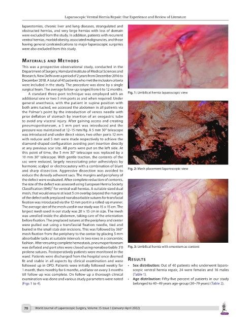
A standard three-port technique was employed with an Fig. 1: Umbilical hernia laparoscopic view
additional one or two 5 mm ports as and when required. Under
general anesthesia, with the patient in supine position with
both arms tucked, we accessed the abdomen in all patients via
the Palmar’s point by the introduction of veress needle with
prior deflation of stomach by insertion of an orogastric tube
to avoid any visceral injury. After gaining access and creating
pneumoperitoneum, a 5 mm port was introduced and the
pressure was maintained at 12–15 mm Hg. A 5 mm 30° telescope
was introduced and under direct vision, two other ports 12 mm
with reducer and 5 mm were made respectively to achieve the
diamond-shaped configuration avoiding port insertion directly
at any previous scar site. All ports were put on the left side. At
this point of time, the 5 mm 30° telescope was replaced by a
10 mm 30° telescope. With gentle traction, the contents of the
sac were reduced, largely necessitating prior adhesiolysis by
harmonic scalpel or electrocautery with a combination of blunt
and sharp dissection. Aggressive dissection was avoided to Fig. 2: Mesh placement laparoscopic view
reduce the densely adherent sacs. The margins and periphery of
the defect were evaluated. After complete reduction of contents,
the size of the defect was assessed using European Hernia Society
7
Classification (EHS) for ventral wall hernias. A suitable sized dual
mesh, that would ensure at least 5 cm overlap beyond the margins
of the defect with preplaced nonabsorbable sutures for transfacial
fixation was introduced via the 12 mm port in a rolled-up manner.
The average size of the mesh used in our study was 15 × 15 cm. The
largest mesh used in our study was 20 × 15 cm in size. The mesh
was unrolled inside the abdomen, taking care of the orientation
before fixation. The preplaced sutures at the periphery and center
were pulled out using a transfascial fixation needle, tied and
buried in the small stab skin incisions. This was followed by 360°
mesh fixation from the periphery to the center by placing 5 mm
absorbable tacks at suitable intervals in two rows in a concentric
fashion. After ensuring complete hemostasis, pneumoperitoneum
was deflated and port sites were closed using nonabsorbable 3’0 Fig. 3: Umbilical hernia with omentum as content
prolene sutures. Postoperatively patients were monitored in the
ward. Patients were discharged from the hospital once deemed
fit and stable in all aspects by clinical examination and were results
followed up in OPD. Patients were initially followed weekly for • Sex distribution: Out of 40 patients who underwent laparo-
1 month, then monthly for 6 months, and later on every 3 months scopic ventral hernia repair, 24 were females and 16 males
till follow-up was complete. On follow-up a thorough clinical (Table 1).
examination was done and various study parameters were noted • Age distribution: Fifty-five percent of patients in our study
(Figs 1 to 4). belonged to 40–49 years age-group (30–79 years) (Table 2).
70 World Journal of Laparoscopic Surgery, Volume 15 Issue 1 (January–April 2022)