
Page 5 - World Journal of Laparoscopic Surgery
P. 5
WJOLS
Comparative Study of Surgical Approaches for Renal Pelvic Stones in a Northern Rural Medical College
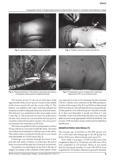
Fig. 2: Landmarks for port placement for left LPL Fig. 3: Position of ports for performing left LPL
Fig. 4: Placement/insertion of DJ stent in renal pelvis and closure Fig. 5: Postoperative scar in a patient who underwent
of pyelotomy being carried out laparoscopically laparoscopic RPL at our medical center
The 1st port of size 1.5 cm was at renal fossa at the was removed as soon as the drainage became minimal
upper border of the erector spinae muscle (in the middle (<20 mL). Stiches were removed on the 10th postopera-
of the lower coastal rib and the coccyx) (Fig. 2). The tive day of the surgery (Fig. 5) X-ray KUB and ultrasound
balloon was inflated with water and kept inflated for KUB were done to rule out retained stone postoperatively.
3 minutes to achieve adequate dissection and hemostasis. All the patients were followed up for 6 months, initially
The 2nd port was established in the renal angle of size at 15 days and thereafter 1 month and then at 3 and
5 mm (Fig. 3). The third port of 5 mm was made above 6 months. At the end of the study, the data were collected
the iliac crest, which was converted into an 8 mm port to and analyzed using appropriate statistical methods. The
insert the cold knife for pelvic incision. The renal pelvis p-value ≤ 0.05 was taken as the cutoff point for statistical
was incised with endoscissor/cold knife. significance.
The stone was grabbed with an endograsper or artery
forcep, whichever was easier to hold the stone. The stone OBSERVATIONS AND RESULTS
was pulled out of renal pelvis and kept near to the ureter. The average age of patients in the RPL group was
The ureteric stent was placed and the pelvis was closed 37.1 ± 12.29 years and average age in the OP group was
with absorbable 4-0 vicryl suture (Fig. 4). 46.66 ± 10.39 years. Male to female ratio was 2.33:1.
Cystoscope was inserted through the lower 5 mm From Table 1, in group I, 112 (40%) of the cases were
port site and under evidence of cystoscope, the pelvic completed within 61 to 70 minutes and 140 cases (50%)
stone was removed through the 10 mm port incision site. were completed in >70 minutes. Hence, it was found
The patient was discharged on the 3rd or 4th day of that the maximum number of cases [140 (50%)] were
surgery according to the condition of the patient. Drain completed in >70 minutes. Whereas in group II, similarly,
World Journal of Laparoscopic Surgery, January-April 2017;10(1):1-7 3