DR VISHWANATH MOGALI MBBS, DNB (Gen.Surg), F.MAS, D.MAS
General and advanced laparoscopic surgeon
Saveetha medical college and hospital Chennai, India
This task analysis is submitted as a part of fulfillment of rules and regulations of world laparoscopy hospital for the award of Fellowship and diploma in minimal access surgery - December 2016 batch
TASK ANALYSIS OF SILS CHOLECYSTECTOMY
INTRODUCTION
Cholecystectomy is the most common major abdominal procedure performed now a day. Management of gall stone disease has progressed through eras of nonsurgical management, laparotomy, mini-laparotomy and now laparoscopic cholecystectomy which is the gold standard treatment for benign diseases of gall bladder.
Laparoscopic surgery is the procedure of choice for most benign gall bladder diseases unless contraindicated. The advantages like earlier return of bowel function, less postoperative pain, good cosmesis, shorter length of hospital stay, earlier return to full activity were appreciated.
As the technology and instruments improved, surgeons modified the procedure from traditional four ports laparoscopy to three ports and then to two ports.
As surgeons and industry continue to push the boundaries of laparoscopic or minimally invasive surgery(MIS), new and controversial approaches such as natural orifice trans-luminal endoscopic surgery (NOTES) and single-incision laparoscopic surgery(SILS) are being explored with the goal of reducing surgical morbidity.
Various natural orifices like mouth (trans-gastric), umbilicus and vagina are being used as portals for surgery. Termed variously as Single Port Access(SPA) surgery, Single Incision Laparoscopic Surgery (SILS) or OnePort Umbilical Surgery (OPUS) or Single Port Incision Less Conventional Equipment Utilizing Surgery (SPICES) or Natural Orifice Trans-umbilical Surgery (NOTUS).
SILS is a novel technique which promises all advantages of minimally invasive surgery with additional benefits like reduced postoperative morbidity, improved cosmesis and patients acceptance.
INDICATIONS:
1. Symptomatic gallstones causing repeated episodes of biliary colic.
2. Mucocele of gallbladder, biliary pancreatitis after ERCP and CBD stone extraction.
3. Cholecystitis - acute calculus/acalculus cholecystitis, chronic cholecystitis.
4. Gall bladder polyp size more than 1cm is also an indication for cholecystectomy.
5. In asymptomatic cholelithiasis indications include diabetics, patients undergoing bariatric surgery, renal transplantation, and those with hemolytic diseases.
CONTRAINDICATIONS:
1. Patients who are not fit for general anesthesia.
2. Significant portal hypertension, uncorrectable coagulopathy.
3. Patients with proven or suspected carcinoma of gallbladder.
4. Relative contraindications include complications of cholecystitis like empyma, gangrenous gall bladder and perforation of gallbladder, morbid obesity, pregnancy and cirrhosis of liver.
TASK ANALYSIS SILS CHOLECYSTECTOMY
1. Preparation and anesthesia
2. Procedural steps
3. Executional steps
1. Preparation and anaesthesia:General anaesthesia with Ryle’s tube for decompression of stomach, Bladder catheterization using 14 or 16 Fr Folay’s catheter.
2. Procedural steps:
I. French position of the patient, painting with betadine and draping.
II. Arrangement of optical instruments and energy sources and instrument tray.
III. Creation of pneumoperitoneum and insertion of SILS port and trocars.
IV. Retraction of fundus and dissection of calots triangle to expose cystic artery and cystic duct.
V. Clipping and division of cystic artery and cystic duct.
VI. Dissection of GB from liver and hemostasis.
VII. Specimen retrival along with removal of trocars and SILS port.
VIII. Closure of fascial layer followed by skin.
3. Executional steps:
SINGLE INCISION LAPAROSCOPIC HOLECYSTECTOMY:
Since SILS procedure is relatively new and in evolution, many techniques happen to be described but no widely accepted standard exists. As the primary benefit of SILS seems to be cosmetic, most agree that the umbilicus may be the preferred incision site.
I. Patient Positioning: Patient can be put within supine, split leg lithotomy, or French position. Most prefer the French position in which the patient’s legs split with slight reverse-Trendelenburg position. Surgeon stands between the patient’s legs and assistant surgeon stands either left or right of the surgeon to hold camera, and second assistant who stands either left or right of the patient. The scrub nurse usually stands on left side of the patient. The camera monitor is placed usually at head end or at right shoulder as in standard laparoscopic cholecystectomy. The light source and insufflator are placed similar to the laparoscopic cholecystectomy positioning.

Figure 1. Position of patient and operation room set up in SILS cholecystectomy.
S-Surgeon stands between patients leg, A-Assistant stands to hold camera.
II. Instruments and Devices: All the instruments and devices mentioned above for laparoscopic cholecystectomy are required along with SILS port and 5mm Apple trocars or trocars with small head. Unlike standard laparoscopy, all the trocars, usually 3 to 4, are crowded into one skin incision. To allow the greater freedom of movement and reduced clashing, a few modified trocars with smaller heads, lower profile and absence of insufflation ports such as Apple trocar (Apple Medical Corporation, Marlborough, Massachusetts) and Ternamian EndoTIPTM (Karl Storz Endoscopy, Tuttlingen, Germany). This allows freedom from the hands while maximizing technique incision. Some surgeons use instruments directly through with no trocars. Purpose-designed ports include multilumen, single-trocar system, such as the R-port (Advanced Surgical Concepts, Wicklow, Ireland), Uni-X single laparoscopic port system, and GelPort (Alexis). Recently, Covidien received FDA clearance to promote its SILS multiple instrument access port.
Flexible or roticulating or articulating instruments are preferable for SILS such as grasper, forceps and clip applicator. Most of surgeons use 10mm clip applicator however 5mm clip applicator if available should be used. The camera scope in SILS cholecystectomy can be 10mm or 5mm with 30 or 45 degree angulation.

Figure 2.SILS kit with port, trocars and roticulating instruments.
III. Incision and port placement: It is advisable to do a formal diagnostic laparoscopy using 10mm vertical incision before the actual SILS incision for the beginners. If the anatomy is clear and less adhesions the umbilical skin incision can be increased to 2cm and fascia, sheath and peritoneum should be divided. Incision can vertical in the umbilicus or ohm (Ω) shape over superior crease of the umbilicus. Then the SILS port should be introduced through the umbilical incision (fig 4) and the gas tube should be connected to create pneumoperitoneum.
IV. Pneumoperitoneum is similar to the laparoscopic cholecystectomy procedure except for, pneumo port in SILS port is separate small tube which connects gas tube outside and enter through the SILS port and opens into abdominal cavity side of SILS port. Once the SILS port is introduced through the umbilical incision the pneumo port should be connected to gas tube and pressures are maintained as in any laparoscopic procedures. Some surgeons use gas evacuator tube which sucks out gas when fumes are build up inside the abdominal cavity due to use of cautery or Harmonic scalpel.
V. SILS port which contains 3 small holes for trocar placement, one 10mm trocar inserted through lower hole, which is used for camera scope. Another 5mm or 10mm trocar is inserted through the upper right hole in SILS port and finally the upper left hole is used for 5mm trocar insertion. Three 5mm trocars (Apple trocars) can be used if 5mm camera scope and 5mm clip applicator are available or only one 10mm or 12mm trocar for camera scope and remaining two 5mm trocars can be used.
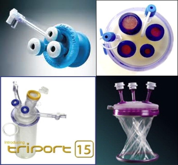
Figure 3.Various SILS ports.a. Covidien SILS port, b. Quad port, c.Triport 15 and d.GelPoint.
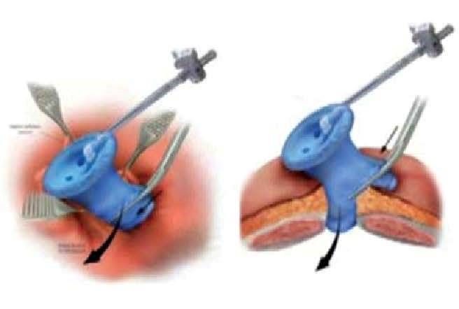
Figure 4.Virtual Picture showing SILS port placement.
VI. Instruments: Once the SILS port is introduced, trocars are placed, and pneumoperitoneum created, the 5mm or 10mm diameter, 30 degree laparoscope inserted through the camera port. Endo Eye coaxial camera(fig 6) can be used to avoid collision of light cable with instruments. The 5mm roticulating grasper is introduced through the upper left trocar and held in surgeon’s left hand. Through the upper right trocar the hook with cautery or Harmonic scalpel™or Maryland dissecting forceps can be used. The 5mm diameter suction cannula, 5mm or 10mm clip applicator, laparoscopic curved scissor, toothed grasper or claw forceps should be kept ready.
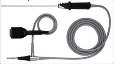
Figure 5. Endo EYE with coaxial light cable.
VII. Gallbladder fundus retraction:This is an important step in SILS cholecystectomy which is not usually done in standard laparoscopic cholecystectomy. The fundus retraction in SILS cholecystectomy can be done using 2-0 ProleneTM (Polypropylene Suture; Ethicon, Johnson & Johnson Intl, Sint-Stevens-Woluwe, Belgium) suture on straight needle which is passed through the abdominal wall under vision at the right mid-clavicular line about two finger breadth above the right costal margin and then passed through the fundus of the gallbladder and back again through the abdominal wall and tied on the outside of the abdominal wall. Gallbladder can be retracted using two sutures, one at fundus and another at Hartmann’s pouch. The fundus retraction can also be done using Veress needle inserted through the sub-xiphoid 2mm incision.
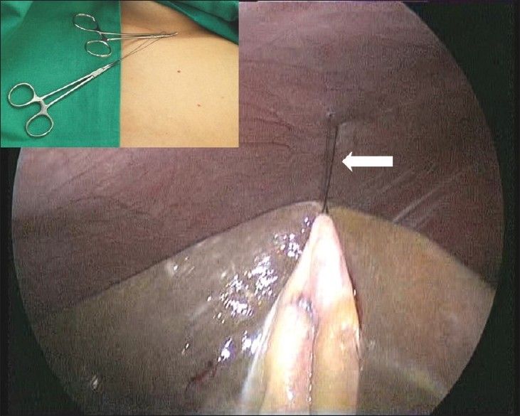
Figure 6.Laparoscopic view of GB fundus traction with external view of suture clip.
VIII. Procedure: After fundus of gallbladder retracted with suture, the remaining steps are similar to the standard laparoscopic cholecystectomy but difference is that the left hand instrument is used to dissect gallbladder while right hand instrument for retraction of Hartman’s pouch. The Hartman’s pouch held with the roticulating grasper and the dissection of triangle of Calot is done using Maryland forceps or hook with cautery or Harmonic scalpel. The posterior peritoneum is divided tofree the Hartman’s pouch. This is followed by further dissection of the anterior and posterior peritoneal leaves overlying the Calot’s triangle. The cystic artery and cystic duct are skeletonized similar to standard laparoscopic cholecystectomy and end point of this dissection is obtaining a “critical view” showing the window between the cystic duct and artery, and between the cystic artery and the liver. These two windows in the Calot’s triangle are dissected well so as to safely observe the tip of the instrument controlling artery and the duct. Then the cystic artery clipped with two medium sized clips using 5mm or 10mm reusable clip applicator and artery is divided using either scissor or Harmonic scalpel. After dividing the artery, the cystic duct clipped with two medium or large sized clips slightly away from cystic to common bile duct junction and one clip on duct close to the gallbladder. Then the cystic duct is divided using curved scissor.
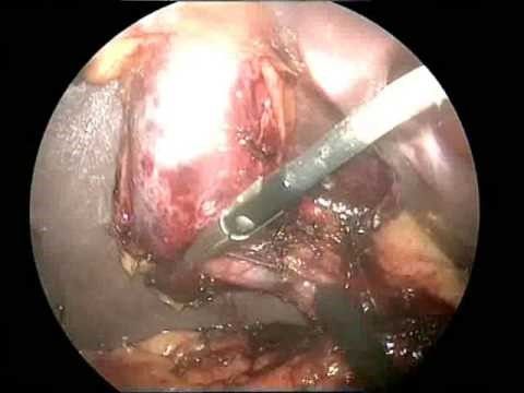
Figure 7.Dissection of Calot’s triangle in SILS.
Once both cystic artery and cystic duct divided safely, the dissection of the gallbladder using hook with cautery or Harmonic scalpel is done by alternating medial and lateral rotation of infundibulum held with roticulating grasper. The gallbladder is separated from the liver and suture applied to retract the fundus is divided. Fundus retraction released by cutting the suture. The gallbladder placed in a sterile plastic bag and kept over the liver waiting for extraction. At this stage the gallbladder fossa is inspected for any bile leak or bleeding, also the cystic duct remnant and cystic artery stump are inspected for bile leak or bleeding. When in doubt a thorough saline wash can be given to gallbladder fossa, and fluid suctioned out meticulously. Any inadvertent spillage of stones are grabbed and placed in the plastic bag containing gallbladder. Once the good hemostasis is ensured, the plastic bag containing gallbladder is removed along with all the instruments and SILS port through the umbilicus.
IX. Closure of the incision: Careful closure of the umbilical incision is mandatory to prevent formation of port site incisional hernia. The edges of the fascial incision are identified, grasped and elevated with Allis forceps or Kocher’s forceps. A non-absorbable suture material like no 1 or 1-0 Prolene or Ethilon can be used for closure of fascial layer in continuous manner or two to three Figure -of-eight manner. After closing the fascia, local anaesthetic infiltration to fascia and skin can be done. The skin is closed with absorbable monofilament suture material by running subcuticular fashion.
Complications of SILS Cholecystectomy:
I. Hemorrage due to injury to cystic artery
II. Bile leak due to CBD or CHD injury.
III. Infection of surgical site.
IV. Incisional hernia.
Difficulties in SILS Cholecystectomy:
Although SILS has many similarities with conventional laparoscopic techniques, there are also a number of differences. Initially, accessing the target organ with multiple instruments and telescope through a single site creates a lot of ergonomic issues like,
I. Triangulation of conventional instruments is difficult
II. As a result clashing or swording of instruments occurs
III. It is difficult to manipulate the instruments as two hands of the surgeon and camera hand of assistant are placed close proximally each other in the SILS port.
To overcome these difficulties, specialized instruments such as Roticulating instruments,endo EYE coaxial telescopes are required and the learning curve to use this technology is higher and it also implies higher technical and financial burden to the person concerned.
ADVANTAGES:
I. Good cosmosis as the umbilicus can completely hide the scar.
II. Early recovery, reduced post operative pain and quality of life which is almost similar to laparoscopic cholecystectomy.
III. Patient’s acceptance is good compared to NOTES and Conventional laparoscopic cholecystectomy.
IV. Specimen retrival is easy through the port.
V. Comined procedures can performed eg; cholecystectomy with appendicectomy or cholecystectomy with hysterectomy.
VI. Surgeon’s domain compared to NOTES which is also performed by physicians.
DISADVANTAGES:
I. Bad ergonomics as base ball-diamond concept not applicable.
II. Difficult with conventional lap instruments, hence need for specialized instruments.
III. Increased cost due use of specialized instruments.
IV. Long learning curve with more stress on surgeons.
V. Increased incidence of surgical site infection and incisional hernia.
Thanks DR VISHWANATH MOGALI